- Home
- Trend
- Weight Loss Strategies
- Acne Tips
- Hair Health Information
- Blemish Removal Tips
- Acne Scar Removal Tips
- Muscle Building Techniques
- Intimate Care Tips
- Postpartum Intimate Care
- Eye Bags Wiki
- Tips for Face Slimming
- Secret of Permanent Hair Removal
- Breast Enlargement Tips
- Cure to Snoring
- Marionette Lines
- Skin-Tightening Secrets
Pigmented lesions are spots or marks that appear on the skin, varying in appearance and significance, which you and I may have noticed some on our skin. These lesions can range from harmless freckles and moles to potentially serious conditions like melanoma. They form due to the accumulation of melanin, the substance responsible for our skin, hair, and eye colour. While many pigmented lesions are benign and pose no health risks, others can be more concerning and may require medical attention.

What Are Pigmented Lesions and Why They Occur

Pigmented lesions refer to areas of the skin that appear darker than the surrounding tissue due to an excess of melanin. These lesions can vary in size, shape, and colour, ranging from light brown to black. The most common types include freckles, moles, and age spots, which are generally benign. However, certain pigmented lesions can evolve into malignant forms, such as melanoma, which is a serious type of skin cancer.
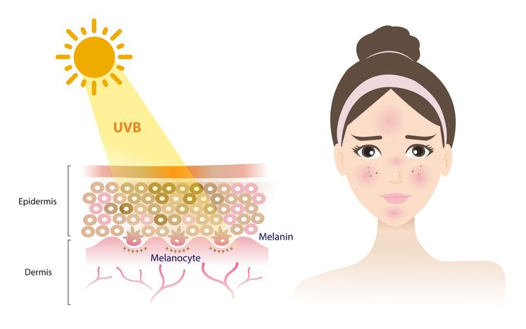
The occurrence of pigmented lesions is influenced by various factors, including genetics, sun exposure, and age. Sun exposure is a major contributor, as ultraviolet (UV) radiation stimulates melanin production in the skin, leading to the formation of lesions like freckles and sunspots. As we age, the cumulative effect of sun exposure can result in age spots, also known as liver spots, which are larger, more defined pigmented areas on the skin.
The Impact of Sun Exposure and Age
Sun exposure is one of the most significant risk factors for the development of pigmented lesions. UV rays from the sun penetrate the skin and damage the DNA within skin cells, prompting an increase in melanin production as a protective response. This process leads to the formation of various types of pigmented lesions, such as sunspots and solar lentigines, which are particularly common in individuals who spend a lot of time outdoors without adequate sun protection.
Age also plays a critical role in the development of pigmented lesions. As the skin ages, its ability to repair itself diminishes, making it more susceptible to the cumulative effects of sun damage. Over time, this can lead to the appearance of age spots and other benign pigmented lesions. However, age also increases the risk of malignant lesions, as the likelihood of skin cells undergoing harmful mutations rises.
Benign vs. Malignant Pigmented Lesions
As we all know, understanding the difference between benign and malignant pigmented lesions is important, as it helps you recognize when to seek medical attention if something seems unusual. Benign lesions, such as freckles, moles, and age spots, are usually harmless and do not require medical intervention unless they change in appearance or cause discomfort. These lesions are generally stable in size and shape, and their colour remains consistent over time.
Malignant lesions, on the other hand, pose a significant health risk and require prompt medical attention. Melanoma, a type of skin cancer, is the most dangerous form of malignant pigmented lesion. It can develop from existing moles or appear as new lesions on the skin. Melanoma is characterised by asymmetry, irregular borders, uneven colour, and changes in size or shape. Other types of skin cancers, such as basal cell carcinoma and squamous cell carcinoma, can also present as pigmented lesions and must be evaluated by a healthcare professional.
免費體驗
PicoCure Pigmentation Removal Treatment
1 Minute Self-Registration
Date should not be before minimal date
Common Conditions: Freckles, Moles, Age Spots vs. Melanoma, Skin Cancers
Freckles, moles, and age spots are examples of benign pigmented lesions that many people develop over their lifetime. Freckles, or ephelides, are small, flat, and usually appear on sun-exposed areas of the skin, especially in individuals with lighter skin tones. Moles, or nevi, are typically brown or black and can be flat or raised. While most moles are benign, some can develop into melanoma. Age spots, also known as liver spots or solar lentigines, are larger pigmented areas that appear on sun-exposed areas of the skin as people age.
Melanoma and other types of skin cancer, such as basal cell carcinoma and squamous cell carcinoma, represent the malignant side of pigmented lesions. Melanoma is particularly dangerous because it can spread to other parts of the body if not detected and treated early. Basal cell carcinoma often appears as a pearly or waxy bump on sun-exposed areas, while squamous cell carcinoma may present as a scaly, red patch or a firm, raised nodule.
Signs and Symptoms That Should Not Be Ignored
Recognizing the warning signs of pigmented lesions is crucial for early detection and treatment, especially when it comes to potentially serious conditions like melanoma. Here are some key signs and symptoms that should prompt immediate medical attention:
1. Asymmetry
One of the hallmark signs of a suspicious lesion is asymmetry. In a benign mole or lesion, both halves usually look alike if you were to draw a line down the middle.
However, if one half of the lesion does not mirror the other, this asymmetry could be an early indicator of melanoma, a serious form of skin cancer. This difference in shape is significant because malignant cells can grow in an uncontrolled and irregular manner, leading to uneven lesions.
2. Border Irregularity
The borders of a benign pigmented lesion are typically smooth and well-defined. On the other hand, lesions with uneven, scalloped, or poorly defined borders should raise concern.
These irregular borders occur because cancerous cells spread in a haphazard fashion, leading to edges that are not well contained. If you notice a lesion with jagged or fuzzy borders, it is important to have it evaluated by a healthcare professional.
3. Colour Variation
Colour is another critical factor in assessing pigmented lesions. While benign moles generally have a single shade of brown, malignant lesions can contain a variety of colours, including shades of brown, black, red, white, and even blue.
This colour variation is concerning because it may indicate different levels of pigment production or bleeding within the lesion, both of which are associated with melanoma. The presence of multiple colours in a single lesion should be checked immediately.
4. Diameter
Size matters when it comes to pigmented lesions. Lesions larger than 6 millimetres in diameter—about the size of a pencil eraser—warrant closer examination. While not all large lesions are malignant, larger size can be a characteristic of more advanced skin cancers.
Early detection is crucial, so any lesion that exceeds this size should be brought to the attention of a dermatologist.
5. Evolution
One of the most critical signs to monitor is evolution. Any changes in the size, shape, colour, or elevation of a lesion, or the appearance of new symptoms such as bleeding, itching, or crusting, are red flags that should not be ignored.
Evolution suggests that the lesion is active, and changes over time could indicate malignancy. It’s essential to monitor existing lesions for any alterations and to report new symptoms to a healthcare provider immediately.
Additional Symptoms to Watch For
While the ABCDEs (Asymmetry, Border, Color, Diameter, and Evolution) are the primary indicators, there are other symptoms that should prompt a visit to a dermatologist. These include:
• Pain or Tenderness: A pigmented lesion that becomes painful or tender is unusual and should be examined.
• Bleeding or Oozing: Any lesion that starts to bleed or ooze without an apparent cause could be indicative of a serious condition.
• Rapid Growth: A lesion that appears to be growing quickly or spreading across the skin is cause for concern.
• New Pigmented Lesions: The sudden appearance of a new pigmented lesion, particularly in an adult, should be assessed to rule out malignancy.
The Role of Dermatologists in Early Diagnosis
If you suspect you have malignant pigmented lesions, dermatologists play a vital role in the early diagnosis of pigmented lesions. They are trained to recognize the subtle differences between benign and malignant lesions and can perform thorough skin examinations to identify areas of concern. Regular check-ups with a dermatologist are especially important for individuals with a family history of skin cancer or those who have numerous moles or other pigmented lesions.
Diagnostic Techniques: Dermoscopy, Biopsy, and Imaging
Several diagnostic techniques are used to evaluate pigmented lesions. Dermoscopy is a non-invasive method that allows dermatologists to examine the skin with a specialised magnifying tool, providing a clearer view of the lesion's structure and pigmentation patterns. This technique helps distinguish between benign and malignant lesions.
If a lesion appears suspicious, a biopsy may be performed. This involves removing a small sample of tissue from the lesion and examining it under a microscope to determine if cancerous cells are present. In some cases, imaging techniques such as reflectance confocal microscopy (RCM) or optical coherence tomography (OCT) may be used to provide detailed images of the skin layers without the need for a biopsy.
Treatment Approaches
Once a pigmented lesion has been diagnosed, the appropriate treatment approach will depend on whether it is benign or malignant, as well as the lesion's size, location, and patient preference.
免費體驗
PicoCure Pigmentation Removal Treatment
1 Minute Self-Registration
Date should not be before minimal date
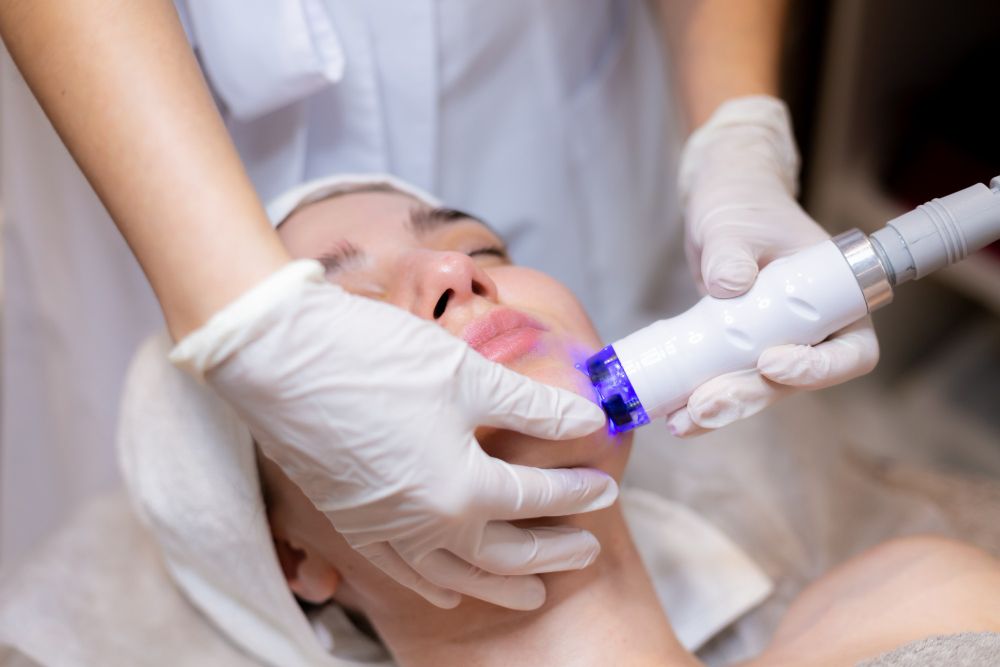
PicoCure Pigmentation Removal Treatment: A Breakthrough in Treating Benign Pigmented Lesions

Malignant lesions can be dangerous, so it’s crucial to have them checked at a hospital, especially if you have a family history of skin cancer or numerous moles or other pigmented lesions. For less severe or benign pigmented lesions, however, PicoCure Pigmentation Removal Treatment can be your solution.
PicoCure Pigmentation Removal Treatment is an advanced laser treatment that has revolutionised the approach to treating pigmented lesions, particularly those that are benign but aesthetically concerning, such as age spots and freckles. The PicoCure laser uses picosecond technology, which delivers ultra-short pulses of energy to target pigment particles within the skin. This rapid energy delivery breaks down the pigment into smaller fragments, which are then naturally eliminated by the body's immune system.
How PicoCure Pigmentation Removal Treatment Works
PicoCure operates on a picosecond scale, delivering energy pulses that are one trillionth of a second long. This allows for highly precise targeting of pigmented lesions without causing significant damage to the surrounding skin. The laser's energy shatters the pigment particles, which are then gradually removed by the body's lymphatic system. This process reduces the appearance of pigmented lesions and results in a more even skin tone.
Treatment's Benefits
• Minimal Downtime: One of the key advantages of PicoCure is its minimal downtime. Patients can typically resume their normal activities shortly after treatment, making it a convenient option for those with busy lifestyles.
• Suitability for All Skin Types: PicoCure is effective on a wide range of skin types, including darker skin tones that are more prone to post-inflammatory hyperpigmentation. This makes it a versatile treatment option for patients with different skin colours.
• Precision: The precision of PicoCure's picosecond technology allows for targeted treatment of pigmented lesions without damaging the surrounding skin. This reduces the risk of scarring and other side effects, which can be a concern with traditional laser treatments.
When to Consider PicoCure Pigmentation Removal Treatment?
PicoCure is an excellent option for patients who have benign pigmented lesions that they wish to remove for cosmetic reasons. It is also suitable for treating stubborn pigmentation issues that have not responded well to other treatments. However, it is essential to consult with a dermatologist to determine if PicoCure is the right treatment for your specific condition.
Get Ahead of Your Skin Lesions
Pigmented lesions are a common feature of the skin, but understanding their causes, potential risks, and treatment options is essential for maintaining skin health. While most pigmented lesions are benign, it is crucial to be aware of the signs of malignancy and seek early diagnosis and treatment when necessary.
Treatments such as PicoCure Pigmentation Removal offer effective, minimally invasive options for managing benign lesions, while surgical and other therapies are available for more serious conditions. Protecting your skin from the sun, staying vigilant about changes, and understanding your genetic risks empower you to take control of your skin health and keep pigmented lesions at bay!
免費體驗
PicoCure Pigmentation Removal Treatment
1 Minute Self-Registration
Date should not be before minimal date
FAQ

1. What exactly are pigmented lesions, and how do they manifest on pigmented skin?
Pigmented lesions are areas of the skin where melanin, the pigment responsible for skin colour, is concentrated. These lesions can range from benign conditions like moles and keratosis to more serious issues like melanoma. On pigmented skin, these lesions may be more challenging to detect due to the natural variation in skin tone.
2. How effective is laser therapy in treating both pigmented and vascular lesions?
Laser therapy is highly effective in treating various pigmented and vascular lesions. For pigmented lesions, lasers target melanin, breaking down the pigment and reducing the appearance of spots. For vascular lesions, lasers work by targeting the blood vessels beneath the skin, helping to diminish the lesion's appearance. Different types of lasers may be used depending on the lesion’s colour and size.
3. How do melanocytes contribute to the formation of pigmented lesions?
Melanocytes are the cells responsible for producing melanin, the substance that gives our skin its colour. When melanocytes produce too much melanin in a concentrated area, it leads to the formation of pigmented lesions, such as freckles, moles, or keratoses. An article on skin health might explore how these cells behave differently in various skin conditions.
4. What distinguishes seborrheic keratosis from other types of skin lesions in adults?
Seborrheic keratosis is a common, non-cancerous skin lesion that appears as a brown, black, or light tan growth on the face, chest, shoulders, or back. Unlike other pigmented lesions, which may arise from sun exposure or other factors, seborrheic keratoses are primarily due to age and are more common in older adults. They are benign and typically do not require treatment unless for cosmetic reasons.
5. Are the substances and methods used in laser therapy for pigmented lesions safe?
Laser therapy for pigmented lesions is generally safe when performed by qualified professionals. The lasers used in these treatments are designed to target specific pigments or vascular structures in the skin, minimising damage to surrounding tissues. However, it’s important to discuss any concerns with your dermatologist, especially if you have sensitive or pigmented skin, to ensure the treatment is appropriate for your condition.